
Unraveling the Connection: Uterine Anomalies in Infertility
Infertility is a complex issue that affects millions of couples worldwide. While various factors can contribute to infertility, uterine anomalies have emerged as a significant cause. These structural abnormalities in the uterus can disrupt the implantation of a fertilized egg, leading to difficulties in achieving pregnancy. In this blog post, we will explore the different types of uterine anomalies and their impact on fertility, diagnosis methods, treatment options, and the hope they bring to couples longing to conceive.
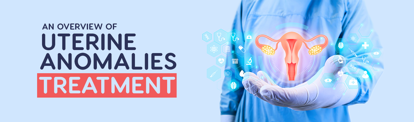
Understanding Uterine Anomalies
Uterine anomalies, also known as uterine abnormalities or congenital uterine malformations, are structural irregularities or variations in the shape, size, or positioning of the uterus. These anomalies occur during the development of the female reproductive system in the womb, leading to differences in the normal structure of the uterus. Uterine anomalies can range from minor variations to more severe malformations that can affect fertility and pregnancy outcomes.
These anomalies can involve different parts of the uterus, including the shape of the uterine cavity, the thickness of the uterine walls, or the presence of abnormal structures within the uterus.
Types of Uterine Anomalies
There are various types of uterine anomalies or uterine malformations, each characterized by unique structural abnormalities. Here are the different types of uterine anomalies:
Uterine Septum:
1. A uterine septum occurs when a band of tissue, called a septum, divides the uterine cavity partially or completely.
2. It is the most common uterine anomaly, accounting for about 35% of all uterine malformations.
3. The septum can vary in size and location, ranging from a small partition to a complete division of the uterus into two separate cavities.
4. This anomaly can interfere with implantation and increase the risk of recurrent miscarriages, preterm birth, and malpresentation of the fetus.

Unicornuate Uterus:
1. A unicornuate uterus is characterized by the absence or underdevelopment of one side of the uterus.
2. This results in a smaller than usual uterus, with a single fallopian tube and ovary on the functional side.
3. It occurs due to the incomplete development or absence of one of the Müllerian ducts during fetal development.
4. Women with a unicornuate uterus may have a higher risk of infertility, miscarriages, and complications during pregnancy, such as preterm labor and breech presentation.
Bicornuate Uterus:
1. A bicornuate uterus is divided into two sections, giving it a heart-shaped appearance.
2. It occurs when the fusion of the Müllerian ducts is incomplete during fetal development.
3. The degree of division can vary, with a mild form having a slight indentation at the top of the uterus and a more severe form having a deep division.
4. This anomaly may increase the risk of recurrent miscarriages, preterm birth, and malposition of the fetus during pregnancy.
Didelphys Uterus:
1. A didelphys uterus, also known as a double uterus, occurs when the uterus develops as two separate structures instead of merging together.
2. Each uterus has its own cervix, and sometimes there can be duplication of the vagina as well.
3. Women with a didelphys uterus may experience difficulties in conceiving, an increased risk of miscarriages, and a higher likelihood of preterm birth.
Arcuate Uterus:
1. An arcuate uterus is characterized by a minor indentation or dip in the upper part of the uterus.
2. It is the mildest form of a uterine anomaly and usually does not significantly impact fertility or pregnancy outcomes.
3. However, in some cases, it may slightly increase the risk of preterm birth.
Other Rare Anomalies:
1. There are other rarer types of uterine anomalies, such as septate cervix (a band of tissue in the cervix) and rudimentary horns (underdeveloped uterine structures).
2. These anomalies can also affect fertility and pregnancy outcomes, requiring specific management approaches.
Diagnosis of Uterine Anomalies
Diagnosing uterine anomalies involves a thorough evaluation by a healthcare professional specializing in reproductive medicine or gynecology. The diagnostic process may include the following steps:
Medical History:
1. The healthcare provider will review your medical history, including any previous pregnancies, miscarriages, or reproductive health concerns.
2. They will ask about your menstrual cycle regularity, symptoms you may be experiencing, and any relevant family history of uterine anomalies or reproductive issues.
Physical Examination:
1. A pelvic examination will be conducted to assess the size, shape, and position of your uterus.
2. The healthcare provider will also examine the cervix and other pelvic structures to check for any visible abnormalities.
Imaging Techniques:
1. Ultrasound: Transvaginal ultrasound is commonly used to evaluate the uterus and detect structural abnormalities. A specialized ultrasound probe is inserted into the vagina to obtain detailed images of the uterus and its internal structures.
2. Hysterosalpingography: This imaging procedure involves injecting a contrast dye into the uterus and fallopian tubes, followed by X-rays. It can help identify uterine abnormalities, such as a uterine septum or bicornuate uterus, as well as assess the patency of the fallopian tubes.
Magnetic Resonance Imaging (MRI):
1. MRI provides more detailed imaging of the uterus and can help identify and classify different types of uterine anomalies.
2. It is particularly useful in cases where a more comprehensive evaluation is required or when the results from ultrasound or hysterosalpingography are inconclusive.
Hysteroscopy:
1. Hysteroscopy is a minimally invasive procedure in which a thin, lighted tube (hysteroscope) is inserted through the vagina and cervix to visualize the inside of the uterus.
2. It allows direct visualization of the uterine cavity and any structural abnormalities. Hysteroscopy can be combined with diagnostic techniques like saline infusion sonography (SIS) to improve the accuracy of the assessment.
Additional Tests:
1. In certain cases, additional tests may be recommended to evaluate specific aspects of uterine function or to assess associated conditions.
2. These tests may include hormonal evaluations, genetic testing, or specialized assessments of the endometrium (the lining of the uterus).
Accurate diagnosis of uterine anomalies is crucial for appropriate treatment planning and management. A comprehensive evaluation involving medical history, physical examination, and imaging techniques helps healthcare providers determine the type and severity of the anomaly, which guides the selection of the most suitable treatment options for each individual patient.
Treatment Options
The treatment options for uterine anomalies depend on the specific type and severity of the anomaly, as well as individual factors such as a woman's reproductive goals, overall health, and fertility status. Here are some common treatment approaches:
Surgical Correction:
1. Surgical intervention may be recommended for certain uterine anomalies, particularly those that significantly affect fertility or pregnancy outcomes.
2. Hysteroscopic Surgery: In cases of uterine septum or certain intrauterine adhesions, hysteroscopic surgery can be performed. A hysteroscope is inserted through the vagina and cervix to remove the septum or adhesions, thus restoring a more normal uterine cavity.
3. Metroplasty: For anomalies like bicornuate uterus or uterine didelphys, metroplasty is a surgical procedure to correct the structural abnormality. It involves reconstructing or reshaping the uterus to create a more functional and normally shaped uterine cavity.
4. Surgical correction can improve the chances of successful pregnancy and reduce the risk of complications such as miscarriage or preterm birth.
Assisted Reproductive Technologies (ART):
1. In some cases, uterine anomalies may not be amenable to surgical correction or may still pose challenges for conception and pregnancy even after surgery.
2. In vitro fertilization (IVF): IVF is an ART procedure where eggs are retrieved from the ovaries, fertilized with sperm in a laboratory, and the resulting embryos are transferred to the uterus.
3. IVF bypasses the structural abnormalities of the uterus, allowing for successful implantation and pregnancy.
4. Additional techniques such as embryo transfer guidance (using ultrasound or fluoroscopy) may be employed to optimize the chances of successful implantation.
Supportive Measures:
1. In cases where surgical correction or ART is not feasible or desired, supportive measures may be employed to optimize fertility and pregnancy outcomes.
2. Hormonal support: Hormonal medications, such as progesterone supplementation, may be prescribed to support the uterine lining and promote embryo implantation.
3. Monitoring and timely intervention: Regular monitoring during pregnancy, including ultrasound examinations and prenatal care, is essential to detect and manage any potential complications associated with uterine anomalies.
The Hope for Couples
Discovering a uterine anomaly may initially bring a sense of despair to couples hoping to conceive. However, it is essential to remember that advancements in medical science and reproductive technologies offer hope. With accurate diagnosis and appropriate treatment, many couples have successfully achieved pregnancy and brought their dreams of parenthood to fruition.
Conclusion
Uterine anomalies can significantly impact a couple's ability to conceive and carry a pregnancy to term. However, with advancements in medical technology and the expertise of fertility specialists, these challenges can be overcome. Whether through surgical intervention or assisted reproductive technologies, the path to parenthood is not closed for couples facing uterine anomalies. By seeking professional help and exploring the available treatment options, many couples have been able to realize their dreams of having a child and building a family.
